On this page
- Who is at Risk?
- Definition of colon and rectal cancer
- What is the colon?
- What causes colorectal cancer?
- What are the symptoms of colorectal cancer?
- How is colorectal cancer diagnosed?
- How does cancer spread?
- How is colorectal cancer staged?
- How is colorectal cancer treated?
- What are the chances of recovery?

Definition of colon and rectal cancer
Definition of colon cancer
Cancer that forms in the tissues of the colon (the longest part of the large intestine). Most colon cancers are adenocarcinomas (cancers that begin in cells that make and release mucus and other fluids).
Definition of rectal cancer
Cancer that forms in the tissues of the rectum (the last several inches of the large intestine closest to the anus).
As these two cancer have similar risk factors and methods of detection they are often collectively referred to colorectal cancer.
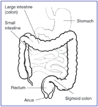
What is the colon?

What causes colorectal cancer?
Age and health history can affect the risk of developing colon cancer.
Anything that increases your chance of getting a disease is called a risk factor. Having a risk factor does not mean that you will get cancer; not having risk factors doesn’t mean that you will not get cancer. People who think they may be at risk should discuss this with their doctor. Risk factors include the following:
- Age 45 or older.
- Other family members with cancer of the colon or rectum.
- A personal history of cancer of the colon, rectum, ovary, endometrium, or breast.
- A history of polyps in the colon.
- A history of ulcerative colitis (ulcers in the lining of the large intestine) or Crohn’s disease.
- Certain hereditary conditions, such as familial adenomatous polyposis (FAP) and hereditary nonpolyposis colon cancer (HNPCC; Lynch Syndrome).
What are the symptoms of colorectal cancer?
Possible signs of colon cancer include a change in bowel habits or blood in the stool.
These and other symptoms may be caused by colon cancer. Other conditions may cause the same symptoms. A doctor should be consulted if any of the following problems occur:
- A change in bowel habits.
- Blood (either bright red or very dark) in the stool.
- Diarrhea, constipation, or feeling that the bowel does not empty completely.
- Stools that are narrower than usual.
- Frequent gas pains, bloating, fullness, or cramps.
- Weight loss for no known reason.
- Feeling very tired.
- Vomiting.
How is colorectal cancer diagnosed?
 The following tests and procedures may be used:
- Fecal occult blood test: A test to check stool (solid waste) for trace amounts of blood. Small samples of stool are placed on special cards and returned to the doctor or laboratory for testing.
- Digital rectal exam: An exam of the rectum. The doctor or nurse inserts a lubricated, gloved finger into the rectum to feel for lumps or anything else that seems unusual.
- Barium enema: A series of x-rays of the colon and rectum. A liquid that contains barium (a silver-white metallic compound) is put into the rectum. The barium coats the lower gastrointestinal tract and x-rays are taken. This procedure is also called a lower GI series.
- Sigmoidoscopy: A procedure to look inside the rectum and sigmoid (lower) colon for polyps, abnormal areas, or cancer. A sigmoidoscope is inserted through the rectum into the sigmoid colon. A sigmoidoscope is a thin, tube-like instrument with a light and a lens for viewing. It may also have a tool to remove polyps or tissue samples, which are checked under a microscope for signs of cancer.
- Colonoscopy: A procedure to look inside the rectum and colon for polyps, abnormal areas, or cancer. A colonoscope is inserted through the rectum into the colon. A colonoscope is a thin, tube-like instrument with a light and a lens for viewing. It may also have a tool to remove polyps or tissue samples (biopsy), which are checked under a microscope for signs of cancer.
- Virtual colonoscopy: A procedure that uses a series of x-rays called computed tomography to make a series of pictures of the colon. A computer puts the pictures together to create detailed images that may show polyps and anything else that seems unusual on the inside surface of the colon. This test is also called colonography or CT colonography.
How does cancer spread?
The three ways that cancer spreads in the body are:
- Through tissue. Cancer invades the surrounding normal tissue.
- Through the lymph system. Cancer invades the lymph system and travels through the lymph vessels to other places in the body.
- Through the blood.
When cancer cells break away from the primary (original) tumor and travel through the lymph or blood to other places in the body, another (secondary) tumor may form. This process is called metastasis. The secondary (metastatic) tumor is the same type of cancer as the primary tumor. For example, if colon cancer spreads to the liver, the cancer cells in the liver are actually colon cancer cells. The disease is metastatic colon cancer, not liver cancer.
How is colorectal cancer staged?
The process used to find out if cancer has spread within the colon or to other parts of the body is called staging. The information gathered from the staging process determines the stage of the disease. It is important to know the stage in order to plan treatment. The following tests and procedures may be used in the staging process:
- CT scan (CAT scan): A procedure that makes a series of detailed pictures of areas inside the body, taken from different angles. The pictures are made by a computer linked to an x-ray machine. A dye may be injected into a vein or swallowed to help the organs or tissues show up more clearly. A CT scan is primary performed to look for metastatic disease.
- Endoscopic ultrasound (EUS): Used to help stage rectal cancer, EUS can accurately gauge the depth the tumor has invaded into the wall of the rectum. Malignant appearing lymph nodes can also be identified by EUS. Performing EUS for rectal cancer helps physicians decided whether chemotherapy and radiation therapy should be given prior to surgery to help shrink the tumor before removing it.
- MRI (magnetic resonance imaging): A procedure that uses a magnet, radio waves, and a computer to make a series of detailed pictures of areas inside the colon. A substance called gadolinium is injected into the patient through a vein. The gadolinium collects around the cancer cells so they show up brighter in the picture. An MRI may help identify metastatic disease in the liver.
- Surgery: The most accurate way to stage a tumor of the colon or rectum is to examine the tumor under the microscope once removed from the body.
How is colorectal cancer treated?
Treatment options depend on the following.
- The stage of the cancer.
- Whether the cancer has recurred.
- The patient’s general health.
Surgery
Surgery (removing the cancer in an operation) is the most common treatment for all stages of colorectal cancer. A doctor may remove the cancer using one of the following types of surgery:
- Resection: For most colon and some rectal cancers a surgeon will perform a partial colectomy (removing the cancer and a small amount of healthy tissue around it). The doctor may then perform an anastomosis (sewing the healthy parts of the colon together). The doctor will also usually remove lymph nodes near the colon and examine them under a microscope to see whether they contain cancer.
- Resection and colostomy: If the doctor is not able to sew the 2 ends of the colon back together, a stoma (an opening) is made on the outside of the body for waste to pass through. This procedure is called a colostomy. A bag is placed around the stoma to collect the waste. Sometimes the colostomy is needed only until the lower colon has healed, and then it can be reversed. If the doctor needs to remove the entire lower colon, however, the colostomy may be permanent.
Even if the doctor removes all the cancer that can be seen at the time of the operation, some patients may be given chemotherapy or radiation therapy after surgery to kill any cancer cells that are left. Treatment given after the surgery, to increase the chances of a cure, is called adjuvant therapy.
Chemotherapy
Chemotherapy is a cancer treatment that uses drugs to stop the growth of cancer cells, either by killing the cells or by stopping them from dividing.
- Systemic chemotherapy – When chemotherapy is taken by mouth or injected into a vein or muscle, the drugs enter the bloodstream and can reach cancer cells throughout the body.
- Chemoembolization may be used to treat cancer that has spread to the liver. This involves injecting anticancer drugs directly into the artery entering the liver. The liver’s arteries then deliver the drugs throughout the liver. Only a small amount of the drug reaches other parts of the body.
The way the chemotherapy is given depends on the type and stage of the cancer being treated.
Radiation therapy
Radiation therapy is a cancer treatment that uses high-energy x-rays or other types of radiation to kill cancer cells or keep them from growing. External radiation therapy uses a machine outside the body to send radiation toward the cancer.
What are the chances of recovery?
The prognosis (chance of recovery) depends on the following:
- The stage of the cancer (whether the cancer is in the inner lining of the colon only, involves the whole colon, or has spread to other places in the body).
- Whether the cancer has blocked or created a hole in the colon.
- The blood levels of carcinoembryonic antigen (CEA; a substance in the blood that may be increased when cancer is present) before treatment begins.
- Whether the cancer has recurred.
- The patient’s general health.
Reprinted with modifications from the National Cancer Institute’s website.

